
news & culture
.avif)
News

Welcome Ana Daniela Huasupoma Mija!
Ana Daniela successfully applied for an Erasmus+ internship program and joined us for her bachelor thesis. Congratulations!

Happy PhD start Pille-Riin Kurrikoff!
After a successful Master's thesis internship, Pille-Riin is now officially enrolled as a PhD in the lab! Looking forward to exploring the -omics of stress vulnerability together.
.avif)
A Fantastic Team Effort: The Collateral VMH Connectome!
Led by Veronika (voltage imaging) and Chiara (optogenetics), we are thrilled to present the first collaboration between the Calvigioni Lab and the Fuzik Lab, shedding light on the Esr1-positive VMH connectome in defensive behaviors! An exciting start to the year—congratulations to the whole team for this incredible work!

Congratulations on Receiving the 2024 Hjärnfonden Project Grant!
We are incredibly grateful for this support and extend a special thanks to the Team Rynkeby Foundation for joining our mission to advance neuroscience and mental health research!
NeuroArt
The beauty of the Brain
Science and art are deeply connected—both driven by curiosity, creativity, and the pursuit of understanding. Here, we share stunning images of the brain (and occasionally the body) to inspire wonder, curiosity, and share our passion for neuroscience. By making the invisible visible, we aim to convey our excitement for the beauty of scientific discovery and the intricate complexity of the brain.

Projection-labelled neurons in the mouse hippocampus.

High-resolution image of developing cortical axons in the embryonic mouse brain.

Hippocampal synaptic terminals in the mouse brain.

The dente gyrus in the adult mouse brain showing parvalbumin-, cholecystokinin-, and secretagogin-expressing neurons.

High-resolution image of developing cortical axons in the embryonic mouse brain.
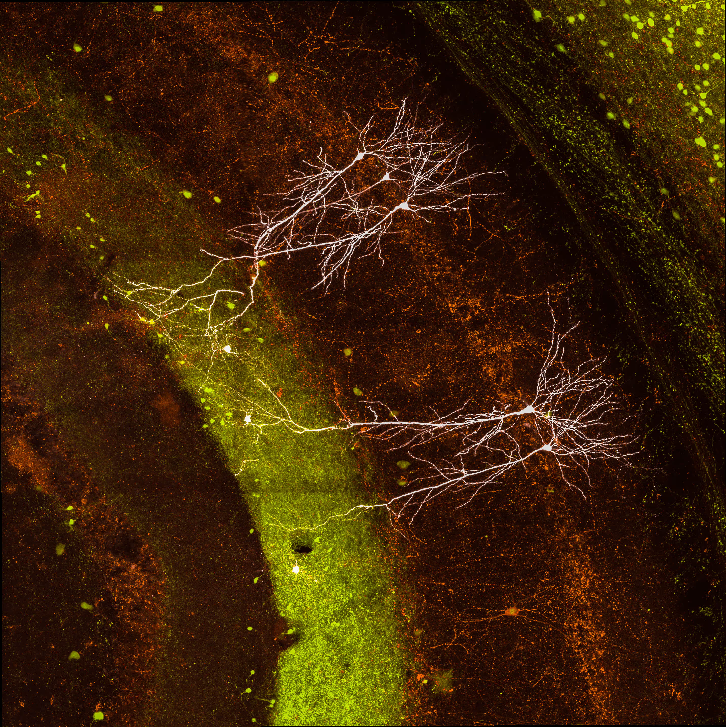
Biocytin filled neurons in the mouse hippocampus.

Heterogenous spines distribution in neighboring pyramidal neurons.

High-resolution image of spines in pyramidal neuron.

A developing neuron in primary cell culture.

The indusium griseum in the adult mouse brain showing parvalbumin-, cholecystokinin-, and secretagogin-expressing neurons.

The dente gyrus in the adult mouse brain showing parvalbumin-, cholecystokinin-, and secretagogin-expressing neurons.

High-resolution image of hippocampal mossy fiber synapses in human.

Pyramidal neurons in the insular cortex labeled by their projection target.

Cross-sections of the musculus gastrocnemius pars interna showing of individual skeletal muscle structure of E18 mouse embryos.

An organotypic slice of an E14.5 embryonic mouse brain shows developing cortical and thalamic axons.

Biocytin reconstruction of a neurogliaform cell, dendrites (yellow) and axon (magenta), and pyramidal neuron (white).

High-resolution image of hippocampal mossy fiber synapses.

Longitudinal sections of the musculus gastrocnemius pars externa showing of individual skeletal muscle structure of P5 mouse.


Lab culture
.avif)

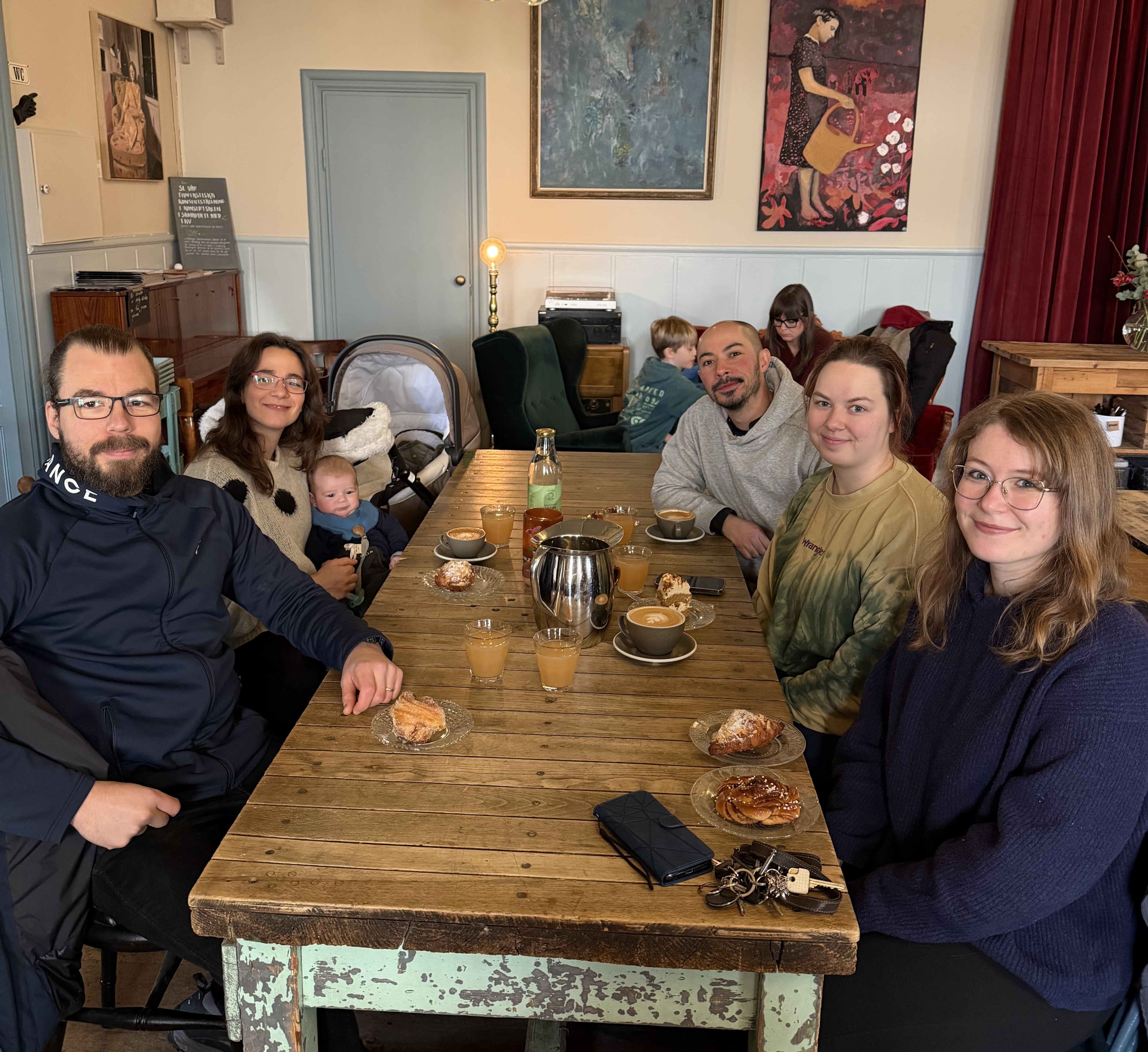
Daniela presents at the StratNeuro Retreat


Center for Social and Affective
Neuroscience (CSAN)
Linköping, Sweden

Department of Neuroscience
Stockholm, Sweden