
news & culture
.avif)
News

SFSG!! Congratulations Daniela!
Daniela is awarded the Swedish Foundation’s Starting Grant, SFSG, from Ragnar Söderbergs stiftelse.The SFSG targets researchers, based in Sweden, who received the highest score on their ERC Starting Grant application but were not funded due to the limited budget of ERC. Five researchers receive funding this year.
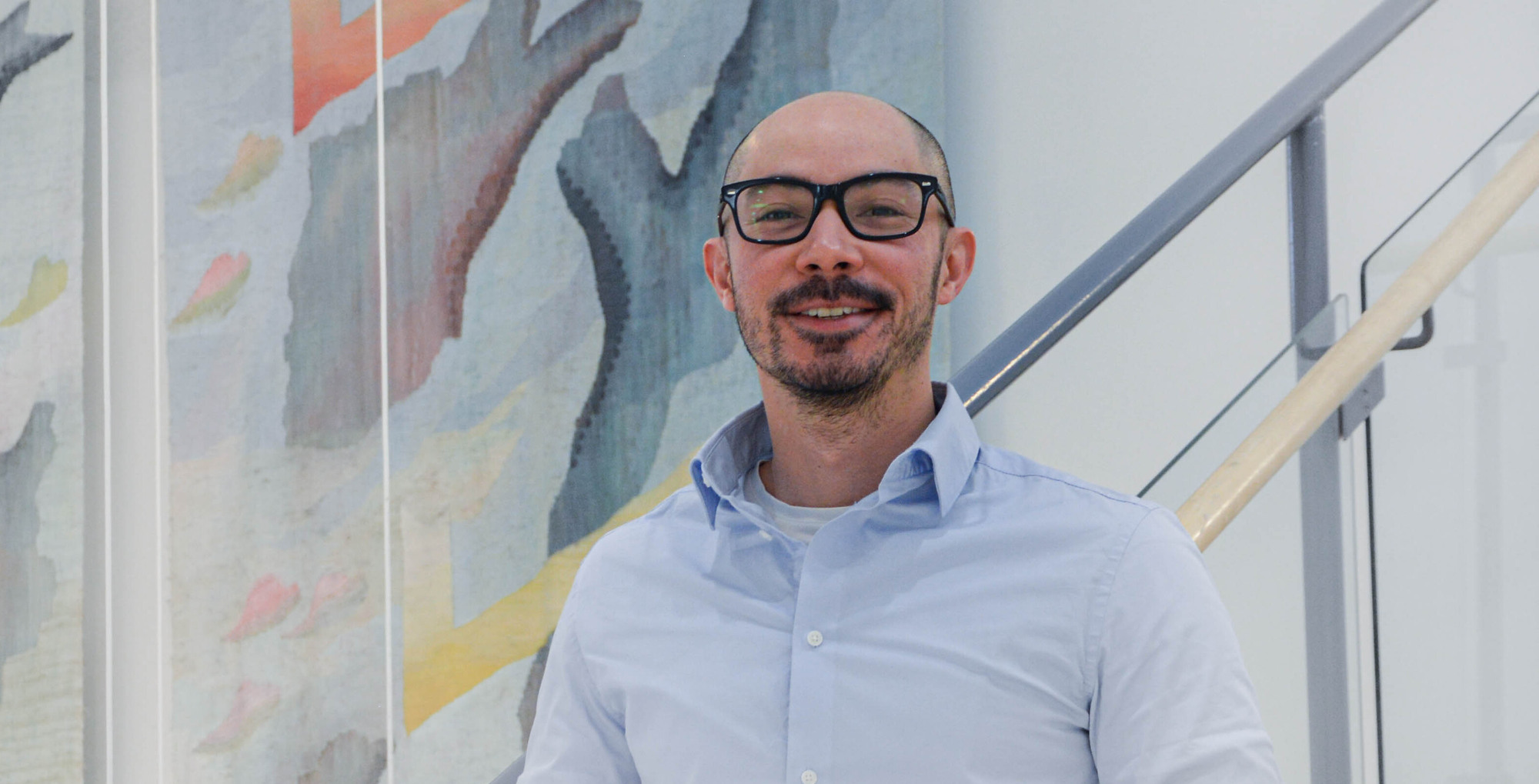
Welcome Sailendrakumar!
Welcome Kumar to Sweden and to our lab!
Kumar joins the lab as postdoctoral fellow, bringing his expertise in patch clamp electrophysiology and insular networks. Juan is the first electrophysiologist of the team!!
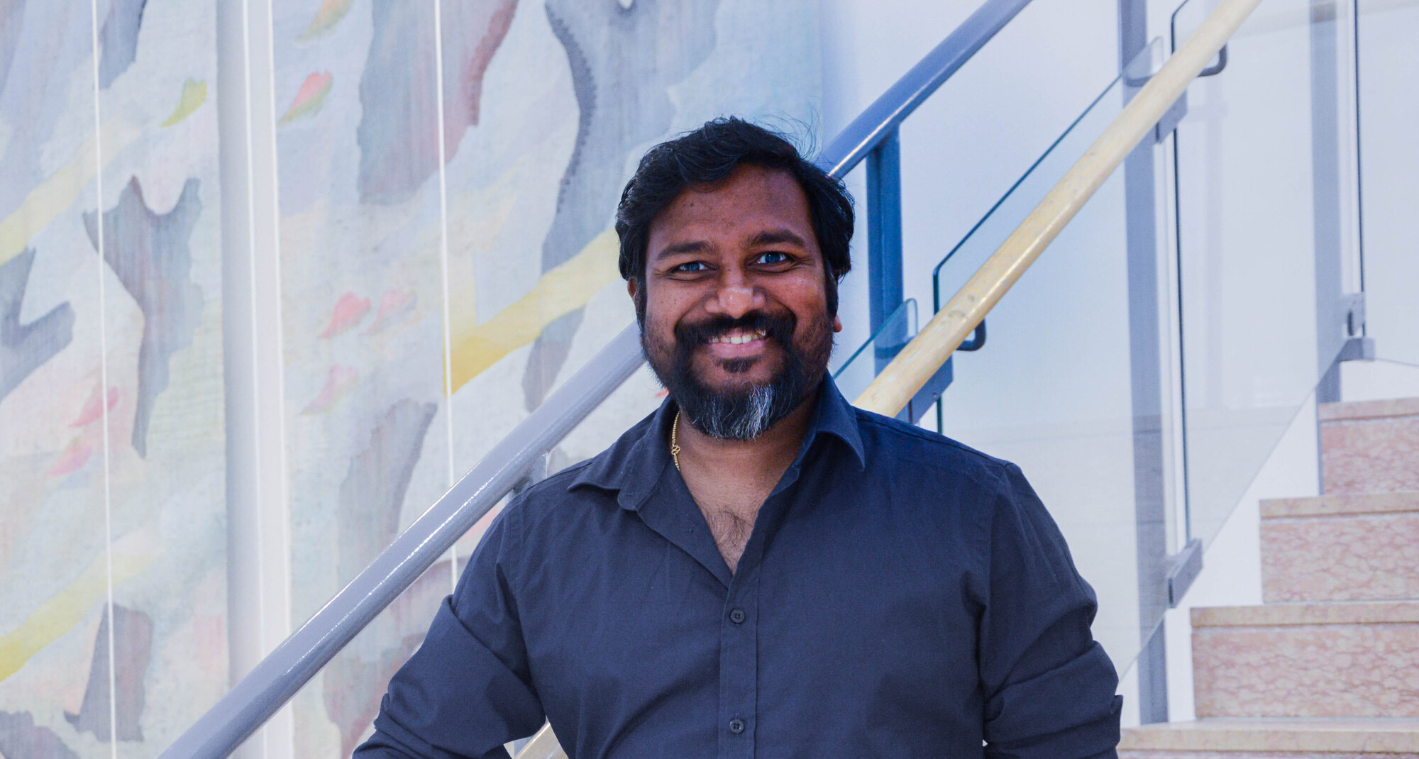
Welcome Juan Martin Uehara!
Welcome Juan to Sweden and to our lab!
Juan joins the lab as postdoctoral fellow, bringing his expertise in social behaviors and freely moving assays development. Juan is the first postdoctoral fellow to join the lab at Linköping University
Best poster award to Pille-Riin Kurrikoff!
Congratulation to Pille-Riin for receiving the best poster award at the 10 years CSAN retreat!
NeuroArt
The beauty of the Brain
Science and art are deeply connected—both driven by curiosity, creativity, and the pursuit of understanding. Here, we share stunning images of the brain (and occasionally the body) to inspire wonder, curiosity, and share our passion for neuroscience. By making the invisible visible, we aim to convey our excitement for the beauty of scientific discovery and the intricate complexity of the brain.

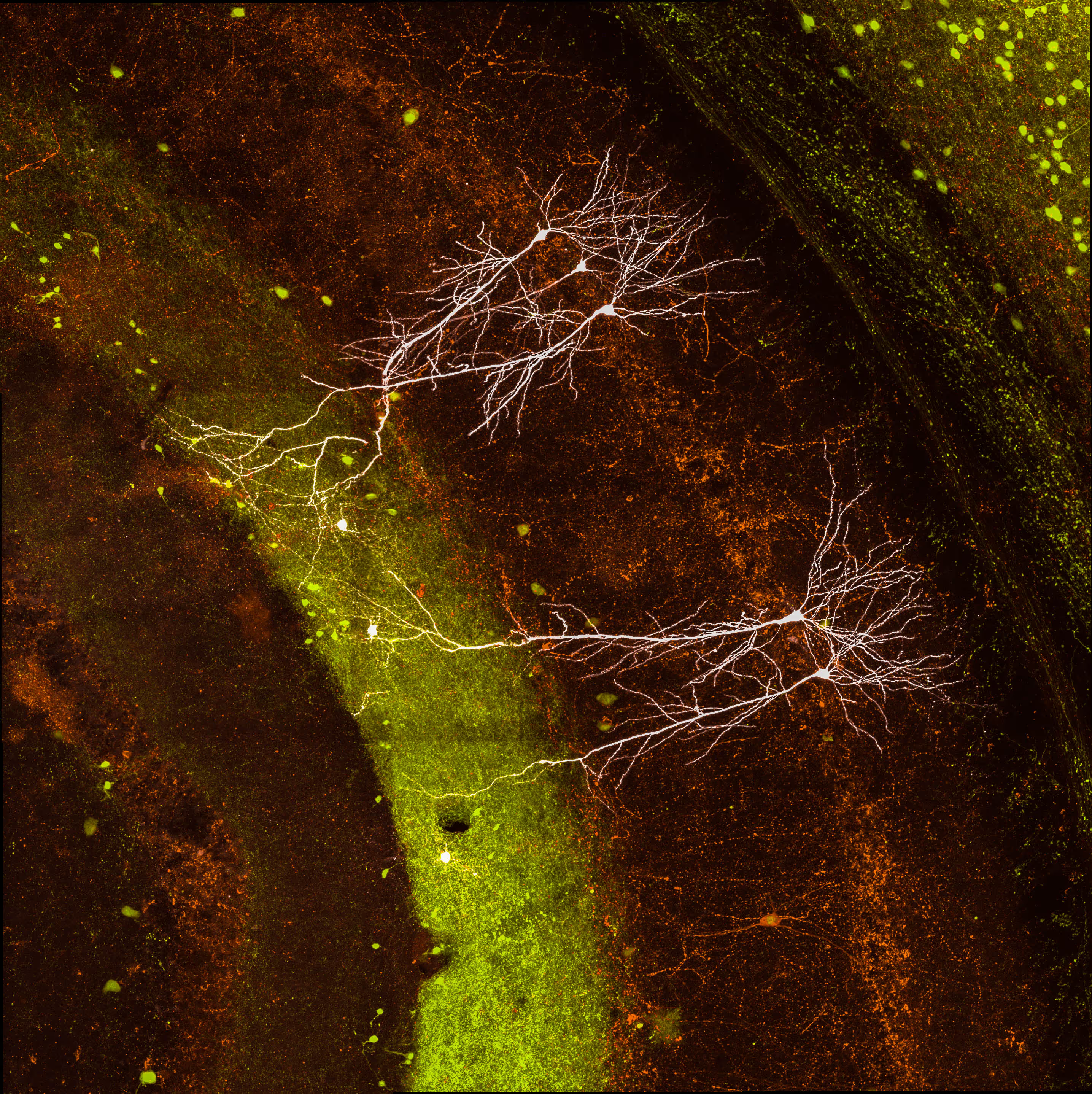
Projection-labelled neurons in the mouse hippocampus.

High-resolution image of developing cortical axons in the embryonic mouse brain.

Hippocampal synaptic terminals in the mouse brain.

The dente gyrus in the adult mouse brain showing parvalbumin-, cholecystokinin-, and secretagogin-expressing neurons.

High-resolution image of developing cortical axons in the embryonic mouse brain.
Biocytin filled neurons in the mouse hippocampus.

Heterogenous spines distribution in neighboring pyramidal neurons.

High-resolution image of spines in pyramidal neuron.

A developing neuron in primary cell culture.

The indusium griseum in the adult mouse brain showing parvalbumin-, cholecystokinin-, and secretagogin-expressing neurons.

The dente gyrus in the adult mouse brain showing parvalbumin-, cholecystokinin-, and secretagogin-expressing neurons.

High-resolution image of hippocampal mossy fiber synapses in human.

Pyramidal neurons in the insular cortex labeled by their projection target.

Cross-sections of the musculus gastrocnemius pars interna showing of individual skeletal muscle structure of E18 mouse embryos.

An organotypic slice of an E14.5 embryonic mouse brain shows developing cortical and thalamic axons.

Biocytin reconstruction of a neurogliaform cell, dendrites (yellow) and axon (magenta), and pyramidal neuron (white).

High-resolution image of hippocampal mossy fiber synapses.

Longitudinal sections of the musculus gastrocnemius pars externa showing of individual skeletal muscle structure of P5 mouse.


Lab culture
.avif)

Daniela presents at the StratNeuro Retreat


Center for Social and Affective
Neuroscience (CSAN)
Linköping, Sweden

Department of Neuroscience
Stockholm, Sweden